











Cardiac ultrasound examination
4.300
RSD
Ultrasound Examination of the Heart | Polyclinic Belgrade
Heart Ultrasound – Polyclinic Belgrade. Echocardiography and assessment of heart function performed by an experienced cardiologist. GREAT PRICE✓ Book your appointment!
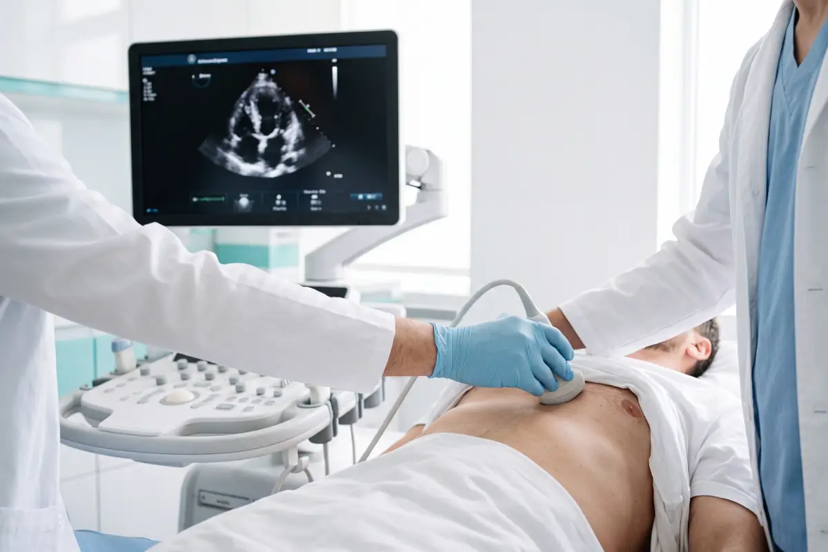
Ultrasound examination of the heart (echocardiography) is a non-invasive and painless diagnostic method that allows for a detailed depiction of the structure and function of the heart. This examination assesses the heart muscle, heart valves, the size of the heart chambers and blood flow through the heart, which enables the early detection of various heart diseases, the monitoring of their course and the planning of appropriate treatment.


An ultrasound examination of the heart, also known as echocardiography, is a painless, safe, and non-invasive diagnostic method in which ultrasound waves are used to display the structure and function of the heart in real time. This examination allows the physician to assess the work of the heart muscle, the heart valves, the size of the heart chambers, the thickness of the heart walls, and the flow of blood through the heart.
Heart ultrasound plays an important role in the detection, diagnosis, and monitoring of various cardiovascular diseases, such as heart valve diseases, heart failure, congenital and acquired heart defects, heart rhythm disorders, and other diseases of the cardiovascular system.
At Poliklinika Beograd, the ultrasound examination of the heart is performed by experienced specialists using modern ultrasound equipment, which allows a precise assessment of the condition of the heart and the planning of further treatment or monitoring of the patient's health condition.
An ultrasound examination of the heart is most often recommended for:
The examination lasts approximately 20 to 30 minutes, is completely painless, does not use radiation, and in most cases no special preparation of the patient is required.
An ultrasound examination of the heart, also known as echocardiography, is a painless, safe, and non-invasive diagnostic method in which ultrasound waves are used to display the structure and function of the heart in real time. This examination allows the physician to assess the work of the heart muscle, the heart valves, the size of the heart chambers, the thickness of the heart walls, and the flow of blood through the heart.
Heart ultrasound plays an important role in the detection, diagnosis, and monitoring of various cardiovascular diseases, such as heart valve diseases, heart failure, congenital and acquired heart defects, heart rhythm disorders, and other diseases of the cardiovascular system.
At Poliklinika Beograd, the ultrasound examination of the heart is performed by experienced specialists using modern ultrasound equipment, which allows a precise assessment of the condition of the heart and the planning of further treatment or monitoring of the patient's health condition.
An ultrasound examination of the heart is most often recommended for:
The examination lasts approximately 20 to 30 minutes, is completely painless, does not use radiation, and in most cases no special preparation of the patient is required.
An ultrasound examination of the heart is recommended when there are symptoms that may indicate heart disease or disorders of its function. This examination enables a detailed assessment of the heart's function and helps in establishing an accurate diagnosis.
The most common symptoms include:
When these symptoms occur or upon the recommendation of a cardiologist, an ultrasound examination of the heart enables an assessment of the function of the heart muscle, valves and blood flow, as well as the timely detection of possible heart diseases and the determination of an appropriate treatment approach.
Cardiac ultrasound examination

Cash - directly before the procedure
Via a dinar or foreign-currency account
RIA, Western Union or MoneyGram
An ultrasound examination of the heart (echocardiography) is a painless and non-invasive diagnostic method used to assess the structure and function of the heart, the work of the heart muscle, the valves, and the blood flow through the heart.
The examination is recommended for symptoms such as chest pain, shortness of breath, heart palpitations, high blood pressure, dizziness, a heart murmur, or as recommended by a cardiologist to monitor an existing condition.
No. A heart ultrasound is completely painless, does not use radiation, and is considered a safe diagnostic method.
The examination usually takes between 20 and 30 minutes, depending on the reason for the examination and the findings.
In most cases, no special preparation is required. The patient can eat, drink, and take their regular therapy normally, unless the doctor recommends otherwise.
The examination can help in detecting diseases of the heart valves, heart failure, congenital and acquired heart defects, disorders of the heart muscle, and other cardiovascular conditions.
No. An ECG and a heart ultrasound are different diagnostic methods that complement each other. An ECG shows the electrical activity of the heart, while an ultrasound shows its structure and function.
The examination is performed by experienced cardiology specialists using modern ultrasound equipment, which enables precise diagnostics and assessment of the heart's function.
Yes. A heart ultrasound examination does not use ionizing radiation and is considered a safe method for all ages, including pregnant women when there is a medical indication for it.
The findings are most often available immediately after the examination, and the cardiologist interprets them and provides recommendations for further treatment, follow-ups, or additional diagnostics if necessary.
Choosing an institution for an ultrasound examination of the heart is important for a precise assessment of the work of the heart and the timely detection of cardiovascular diseases. At Poliklinika Beograd we provide expert diagnostics, modern ultrasound equipment and an individual approach to every patient.
Our patients place their trust in us because of:
Would you like to book an appointment or consult with us? Contact us via Viber or WhatsApp 24/7.

The best aesthetic surgeons in Serbia and the region.
The most affordable surgery prices.



Links
Services
Services
Our partners
Prijatelji sajta